AdenoZAP Kit

Faster method for constructing replication-deficient adenovirus vectors containing transgenes in the E1 region, only one cloning step in E. coli

  • Transgene insertion in E1 region
  • E3 region: WT or 2.7 kb deletion
  • E4 region: WT or 1.2 kb deletion
  • Fiber: WT Ad5 or hybrid Ad5/35
  • Maximum cargo capacity: 9.6 kb
  • Kit contains: E1 shuttle plasmid, RightZAP DNA, adenovirus DNA used as positive control for virus rescue
$590.00
Cat# :
ZK-001
Size: 
10 reactions

Make your kit

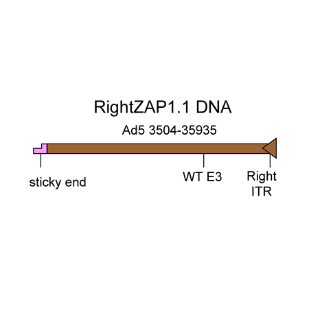
RightZap1.1 DNA   Visit Product Page »

The RightZAP1.1 DNA is a 32.4 kb linear DNA fragment that encompasses bp 3504-right end of the Ad5 genome. The E3 region is intact (WT). The DNA was purified from a cosmid. It is used in combination with the shuttle vectors pZAP1.1 or pZAP1.2 to construct replication-deficient adenoviruses containing transgenes in place of the E1 region of the Ad5 genome. The maximum transgene capacity of the resulting virus is 5.2 kb.

Shuttle plasmid for adenovirus vector construction

pZAP1.1   Visit Product Page »

pZAP1.1 is a 3.5 kb shuttle plasmid designed for inserting expression cassettes in place of the E1 region of the Ad5 genome, in combination with RightZAP1.1 (WT E3), RightZAP1.2 (ΔE3), RightZAP1.3 (ΔE3 + Ad5/35 knob) and other vectors from the AdenoZAP1.0 cloning system. It contains a multiple cloning site downstream from the left ITR and packaging signal of the Ad5 genome. Expression cassettes inserted into this site should contain a promoter, cDNA, and polyA signal. The left arm DNA (which corresponds to the left ITR, packaging signal and expression cassette) can be excised from the vector with either PacI or SwaI on one side, and either PflMI, DraIII, SfiI, BstAPI or AlwNI on the other side.

There are two ways to generate adenovirus vectors with the AdenoZAP system:

1. In vitro ligation and transfection

The AdenoZAP™ cloning system is primarily based on the in vitro ligation of two DNA molecules: the "left arm" and the "right arm".

The left arm is obtained after cloning your gene of interest into a pZAP shuttle plasmid, digesting the resulting plasmid with restriction enzymes and purifying on agarose gel the DNA fragment containing the left ITR, packaging signal and your gene of interest.

The right arm is provided in the kit.  It contains most of the adenovirus genome (from bp 3504 in WT Ad5 to the right end, with several options regarding the status of the E3 and E4 regions, and fiber gene). It originates from a bacterial cosmid rather than from virions, thereby eliminating the danger for a possible viral contamination. It will generate virus only after being ligated to the left arm that contains your expression cassette and the packaging signal.

We have designed the AdenoZAP™ cloning system in such a way that the ligation of both left and right arms is directional. Because of the non-symmetrical nature of the sticky ends generated by excising the left arm from the shuttle plasmid, the ligation of the left and right arms should generate only one type of viral DNA molecule. Consequently, a homogeneous virus population should be generated upon transfection of the helper cells. In addition, such ligation is performed to almost completion in less than an hour using an ordinary T4 DNA ligase.

The ligation product is directly transfected into a helper cell line such as 293 cells. In general, viral plaques appear several days after the transfection.

2. Transfection and in vivo recombination

The second way of creating recombinant adenovirus vectors with the AdenoZAP system is via homologous DNA recombination in helper cells such as 293 cells.  The expression cassettes are cloned into shuttle plasmids pZAP1.2 or pZAP2.2, which contain a 400 bp-long Ad5 DNA sequence downstream from the multiple cloning site.  This Ad5 DNA sequence is part of the pIX gene (bp 3504-3907 in the WT Ad5 genome), and is identical to the left end of the RightZAP DNAs.  The shuttle plasmid is linearized with either PacI or SwaI (whichever is not present in your gene of interest), then transfected into 293 cells together with a RightZAP DNA.  Since both DNA molecules share identical sequences, they can be joined by homologous recombination in 293 cells to create functional recombinant adenovirus vectors.  This approach offers two advantages: (1) the shuttle plasmid needs to be digested with only one restriction enzyme (PacI or SwaI) and (2) there is no need to purify a DNA fragment on agarose gel.  However, the shuttle plasmid must be transfection-grade DNA.

Choosing the AdenoZAP Cloning Kit Suitable for your application.

The AdenoZAP Cloning System exists in different versions and each version can be used in different ways. The following table will help you decide which one is the most appropriate to your application.

Kit Shuttle Plasmid Enzymes available for Left Arm Excision Right Arm E3 Fiber E4 Max Cargo
Left End Right End
AdenoZAP1.1 pZAP1.1 or pZAP1.2 Pac I, Swa I Sfi I, BstAPI, DraIII, AlwNI, PflMI RightZAP1.1 WT Ad5 WT 5.2 kb
AdenoZAP1.2 pZAP1.1 or pZAP1.2 Pac I, Swa I Sfi I, BstAPI, DraIII, AlwNI, Pfl MI RightZAP1.2 Δ 2.7 kb Ad5 WT 7.9 kb
AdenoZAP1.3 pZAP1.1 or pZAP1.2 Pac I, Swa I Sfi I, BstAPI, DraIII, AlwNI, Pfl MI RightZAP1.3 Δ 2.7 kb Ad5/35 WT 8.3 kb
AdenoZAP1.4 pZAP1.1 or pZAP1.2 Pac I, Swa I Sfi I, BstAPI, DraIII, AlwNI, Pfl MI RightZAP1.4 Δ 2.7 kb Ad5/35 Δ 1.2 kb 9.6 kb
AdenoZAP2.1 pZAP2.1 or pZAP2.2 Pac I, Swa I I-SceI RightZAP2.1 WT Ad5 WT 5.2 kb
AdenoZAP2.2 pZAP2.1 or pZAP2.2 Pac I, Swa I I-SceI RightZAP2.2 Δ 2.7 kb Ad5 WT 7.9 kb
  • Only one cloning step in E. coli
    The only cloning you have to perform in E. coli is inserting your expression cassette into a small shuttle vector.
  • No large plasmid (or cosmid) construction and purification

Constructing large plasmids or cosmids is not obvious for inexperienced researchers.  In addition, purifying them in a quality suitable for transfection (i.e. without E. coli chromosomal DNA) is difficult using column chromatography and is better performed on CsCl gradients, which is time-consuming and hazardous.

  • No plasmid midi-prep or maxi-prep necessary

Although using a purified preparation of your shuttle plasmid maximizes the chances of getting your recombinant virus, it is not necessary.  You can use mini-prep DNA: just clean it up after verifying the identity of your clone, digest it with the appropriate enzymes and purify the fragment of interest on agarose gel.  A few micrograms plasmid DNA are usually sufficient to perform the whole procedure.

  • A versatile set of endonucleases available to generate the left arm

Two versions of AdenoZAP cloning system are available.  In AdenoZAP1.0, two 8-base cutters (PacI and SwaI) are available to digest your shuttle vector next to the left ITR, and four 6-base cutters (AlwNI,
BstAPI, DraIII, PflMI) and one 8-base cutter (SfiI) are available to cut your shuttle vector downstream from your expression cassette.  In AdenoZAP2.0, the same 8-base cutters (PacI and SwaI) are available to digest your shuttle vector next to the left ITR, while an 18-base intron-encoded endonuclease (I-SceI) is available to cut the shuttle vector downstream from your expression cassette.  It is therefore likely that this system will be useful in a very large number of applications.

  • Fast

With only one cloning step in E. coli, a DNA digestion, gel purification and a 1-hour ligation before the transfection can be performed, the overall hands-on time is strongly reduced compared to most of the other methods currently available.  If well organized, one can perform the DNA transfection into helper cells only 3 days after starting the cloning of an expression cassette into the shuttle vector and harvest the first virus plaques 3-7 days later.

Adenovirus Expression Vectors

The AdenoZAP cloning system is ideal for constructing replication-deficient adenovirus expression vectors, wherein a transgene expression cassette (promoter + coding sequence + polyA signal) is inserted in place of the E1 region. This is the most popular type of adenovirus vector, also called "first-generation".  The applications include:

  • Transient gene expression in a large variety of cell types and tissues
  • Gene therapy vectors
  • Vaccine vectors

The AdenoZAP cloning system can also be used to construct multi-cistronic adenovirus vectors expressing two or more transgenes.  This can be achieved in multiple ways: (1) by inserting two or more expression cassettes next to each other in the E1 region (pZAP shuttle plasmid); (2) by using IRES-based dicistronic transcriptional cassettes, or (3) by making fusion proteins with self-cleaving P2A peptides.  Another option is to use the AdenoQuick1.0 or AdenoQuick2.0 systems, which allow for inserting expression cassettes at various locations in the adenovirus genome.


  • Ad5 backbone
  • E1 region:
    • 3.1 kb deletion, corresponding to bp 354-3,503 in the Ad5 genome
  • E3 region:
    • WT
    • 2.7 kb BglII deletion, corresponding to bp 28,133-30,818 in the Ad5 genome
  • Fiber:
    • WT
    • Hybrid Ad5/35
  • E4 region:
    • WT
    • 1.2 kb deletion, corresponding to bp 35,319-35,355 in the Ad5 genome and including ORF1-4
  • How do I choose the status of the E3 region: wild-type or deleted?

    In the adenovirus replication cycle, the expression of the E3 region helps the virus evading the host immune system. This region is not essential for virus replication in vitro and therefore can be deleted in order to construct adenoviruses containing longer transgenes, up to 7.7 kb.

    Therefore, if you are using the most common 3.2 kb E1 deletion, and:

    • if your expression cassette (= promoter + coding sequence + polyA signal) is smaller than 5.0 kb, you can use adenovirus vectors with either wild-type or deleted E3 region. 
    • if your expression cassette is larger than 5.0 kb but smaller than 7.7 kb, you must use E1/E3 deleted vectors.
    • if your expression cassette is larger than 7.7 kb, you must consider E1/E3/E4-deleted adenovirus vectors.

    Notes:

    1. In some applications such as oncolytic adenovirus vectors (CRAds), it might be desirable to retain the entire E3 region,  or increase the expression of some E3 products: for instance, the adenovirus “death protein” E3-11.6K, which facilitates the release of viral particles from infected cells, or gp19K, whose constitutive expression reduces the host cytotoxic T cell response against the vector and increases the persistence of transgene expression on its own but possibly not in the context of constitutive expression of the entire E3 region.
    2. The E3 region can also be used as a location to insert a second transgene, independent from the one inserted in the E1 region.
  • How do I calculate the maximum cargo capacity of an adenovirus vector?

    You simply sum up the sizes of the deletions present in your vector and add 1.8 kb. For instance, let us suppose that you are considering using an Ad5 backbone characterized by a 3.2 kb E1 deletion (psn 354-3510) and a 2.7 kb BglII E3 deletion (psn 28133-30818).  Your vector will be able to package 3157 bp (= 3510 - 353)   + 2686 bp (= 30818 - 28132) + 1800 bp = 7.6 kb.

    The additional 1.8 kb is the extra DNA that Ad5 capsids can package in addition to their 35,935 bp-long genome (Bett et al, J. Virol. 1993; 67: 5911-21).

  • What is the maximum cargo capacity for the AdenoQuick and AdenoZAP system?

    For the AdenoQuick1.0 system, the current maximum cargo capacity is achieved with E1/E3/E4-deleted pAd362.  The vector allows for inserting 8.9 kb foreign DNA into the E1 region.

    For the AdenoQuick2.0 system, adenovirus vectors can be constructed, in which up to 11.2 kb foreign DNA can be inserted.  It is done by combining the largest E1 deletion (3157 bp in shuttle plasmid pAd1127-02) with the largest E3 deletion and the hybrid Ad5/35 fiber ( 2686 bp + 756 bp in pAd1129-06), and the largest E4 deletion (2815 bp in shuttle plasmid pAd1130-03), plus the extra 1.8 kb that adenovirus capsids can accomodate in addition to the WT 36 kb genome (Bett et al, 1993. J. Virol. 67: 5911-21).

    For the AdenoZAP system, the current maximum cargo capacity of 9.6 kb is obtained with AdenoZAP1.4.

  • What is the difference between the AdenoZAP1.0 and -2.0 systems?

    Both the AdenoZAP1.0 and AdenoZAP2.0 cloning systems rely on endonucleases that generate non-symmetrical sticky ends with which directional ligation can be performed. The only difference between both systems is the nature of the enzymes available to excise the left arm from the shuttle plasmid pZAP. In pZAP1.1, the left arm can be excised using PacI or SwaI on the left side, and either SfiI, BstAPI, DraIII, AlwNI or PflMI on the right side. PacI and SfiI are 8-base cutters while BstAPI, DraIII, AlwNI and PflMI are 6-base cutters. In pZAP2.1, the left arm can be excised using PacI or SwaI on the left side and I-SceI on the right side. I-SceI is an intron-encoded endonuclease that recognizes an 18-bp-long sequence within which minimal sequence degeneracy is tolerated.

  • I am using the AdenoZAP system. Which enzyme should I use to linearize my shuttle plasmid before transfecting it into helper cells? PacI or SwaI?

    In the AdenoZAP system, you will need to excise from your shuttle plasmid a DNA fragment containing the left ITR, the packaging signal and your expression cassette.  Two enzymes are available to cut on the ITR side: PacI and SwaI.  Make sure that the enzyme you choose does not cut inside your expression cassette.  Since both PacI and SwaI are rare-cutting restriction enzymes that recognize 8 bp-sequences, having both of them in your expression cassette should occur very rarely. 

    If neither PacI nor SwaI cuts inside your transgene, there is really no preference.  Because the PacI site is immediately flanking the start of the adenovirus genome, the DNA ends generated by PacI resemble the most the ends obtained from deproteinised virion DNA and might therefore be more efficient in promoting virus replication. In practice however, no difference in the time needed to recover the virus after DNA transfection into 293 cells is observed between both settings. Virus plaques can appear as early as 4 days after transfecting PacI- or SwaI-digested DNA into 293 cells.

    If one of the two enzymes cuts inside your transgene, use the other one.

    If both SwaI and PacI cut you transgene, eliminate one of them by site-directed mutagenesis, or use another adenovirus construction method.

  • In the AdenoZAP cloning system, what is the difference between pZAP1.1 and pZAP1.2, and also between pZAP2.1 and pZAP2.2?

    pZAP1.1 is the basic AdenoZAP shuttle plasmid.  It contains the adenovirus left ITR, packaging signal, and a multiple cloning site. With this plasmid, you can generate recombinant adenovirus vectors only by ligating the "left arm" to the "right arm" and transfecting the ligation products into 293 cells.  pZAP1.2 is a derivative of pZAP1.1 wherein a 400 bp-long sequence corresponding to bp 3504-3907 in the Ad5 genome was inserted downstream from the multiple cloning site.  With that plasmid, you can generate adenovirus vectors either by ligation and transfection as described above for pZAP1.1, or by transfecting the left arm and the right arm together into 293 cells and have them recombine with each other inside the cell.  The same relationship is true between pZAP2.1 and pZAP2.2.


  • Which facilities are required to work with adenovirus vectors?

    The National Institute of Health has designated adenovirus as Level 2 biological agent.  For most applications, working with adenovirus requires therefore a Biosafety Level 2 (BL2) facility.  The NIH guidelines for research involving recombinant DNA molecules stipulate also that experiments which are likely to either enhance the pathogenicity (e.g. insertion of a host oncogene) or to extend the host range (e.g. introduction of novel control elements) of viral vectors under conditions that permit a productive infection should be performed in BL3 facilities.

    A BL2 laboratory should contain:

    • A warning sign on the entrance door limiting the access to authorized persons only.  The sign should identify the agent, list the name and phone number of the lab director or other responsible person, and indicate any special requirement for entering the lab.
    • A Class II biological safety cabinet.  A Class II cabinet is a ventilated cabinet for personnel and product protection having an open front with inward airflow for personnel protection, and a HEPA filtered mass recirculated air flow for product protection.  The face velocity of the inward flow of air through the full-width open front is 75 feet per minute or greater.  
    • At least one tissue culture incubator dedicated to infected cell cultures.  Another separate incubator is desirable for growing uninfected cells.
    • The minimal equipment to handle adenovirus culture without exiting the BL2 lab (such as centrifuges, microscope…).
    • A sink for hand washing
    • A chemical disinfectant kit or at least a gallon of bleach available for spills

    For more information about guidelines, visit this NIH web page.

  • Which precautions should I take while working with adenovirus?

    Work with adenovirus must be performed in a BL2 lab.  There you must:

    • Always wear a lab coat while in the virus lab.  Before exiting the laboratory for non-laboratory areas (e.g. cafeteria, library, administrative offices…), remove your lab coat and leave it in the laboratory.
    • Avoid skin contamination with the virus.  Always wear gloves (one pair OK, two pairs better for added protection).  Once your gloves have been in contact with infectious material, do not touch common appliances such as telephone or doors handles.  Change your gloves frequently.
    • Keep the lab doors closed while work is in progress.
    • Use mechanical pipetting devices.  Do not pipet by mouth.
    • Decontaminate all work surfaces after you finish your work, and immediately after any spill.  Spray a 10% bleach solution, wipe and spray again a 70% ethanol solution.  For large liquid spills, add directly concentrated bleach to the liquid, leave for at least 5 minutes, and wipe.
    • Perform all procedures with infectious particles in the biosafety cabinet to minimize the exposure of personnel to aerosols.  Minimize the creation of aerosols by pipetting virus cultures and suspension very gently.  Use aerosol-resistant tips for pipetting virus suspensions.  Do not conduct work with infectious materials in open vessels on the open bench. 
    • Use needle-locking syringes or disposable syringe-needle units for the injection or aspiration of infectious fluids.  Extreme care should be used to avoid auto-inoculation and aerosol generation.  Needles should not be bent, sheared, replaced in their sheath or guard or removed from the syringe following use.  The needle and syringe should be decontaminated by pipetting in and out concentrated bleach a few times and then promptly placed in a puncture-resistant container.
    • Decontaminate all contaminated liquid or solid wastes before disposal.  Before starting your virus work, pour some bleach into a beaker.  Rinse all materials (tissue culture dishes, pipets, tips…) that came into contact with adenovirus with 10% bleach inside the hood before discarding them in the Biohazard trash and autoclaving. Place all materials to be decontaminated off-site in a durable leakproof container which is closed before removal. If possible, leave the contaminated materials in contact with bleach for a few hours before autoclaving (e.g. after rinsing your pipets with concentrated bleach inside the hood, soak them in a cylinder containing 10% bleach before autoclaving).
    • Do not leave the BL2 laboratory with live viruses, unless they are in a sealed tube.  Cell cultures transduced with adenoviruses should be inactivated either chemically or biochemically before leaving the BL2 facility.
    • Store your adenovirus preparations at –70 °C in closed containers labeled with Biohazard warning signs.
    • Wash your hands when exiting the laboratory.
AdenoZAP Manual

AdenoZAP Manual

The AdenoZAP Manual contains all the instructions for constructing adenovirus expression vectors using the AdenoZAP system, including:

  • Shuttle plasmid construction
  • Virus Rescue

The manual contains also safety guidelines for handling adenovirus vectors, detailed plasmid maps, and troubleshooting guide.